Having good vision depends on numerous anatomical structures between the orbital bones in one’s face working together properly. There are eight different bones that protect your eyes and the structures within it. For those who don’t have the best vision, it’s likely due to an issue with one of the structures, likely the cornea. There are various treatment options for poor vision, including glasses, contacts, and even Lasik surgery. And to successfully perform these procedures, your surgeon needs an expert knowledge of the anatomy of the eye.
Anatomy of the Eye
Below you can find the parts of the eye. As you will see, the anatomy of the eye has many complex parts.
Eyelids:
Your eyes are covered with lids as a layer of defense from things that may get into your eye. In addition, they keep your eyeballs moist so they can smoothly open and shut. Dense bands of tissue keep your eyelids strong, so they can effectively open and shut.
Orbit:
Your eyeball sits in a protective socket made up of various bones to protect it. Multiple muscles allow the eyes to move up, down, and to the sides.
Sclera:
The muscles attached to the white part of the eye have the name “sclera.” This is the strongest layer of tissue that covers the surface of your eye and is a layer of protection from foreign objects and harmful bacteria.
Tear Structures:
Tears keep your eye moist. And, the tears have three layers. Together the layers comprise the tear film. The lacrimal gland produces the watery part of your tears, and the meibomian gland produces the oily part. The tears drain from your eye through tear ducts.
Cornea:
This is the clear, cone-shaped front portion of the eye where light is focused through. Lasik surgery corrects this portion of the anatomy of the eye.
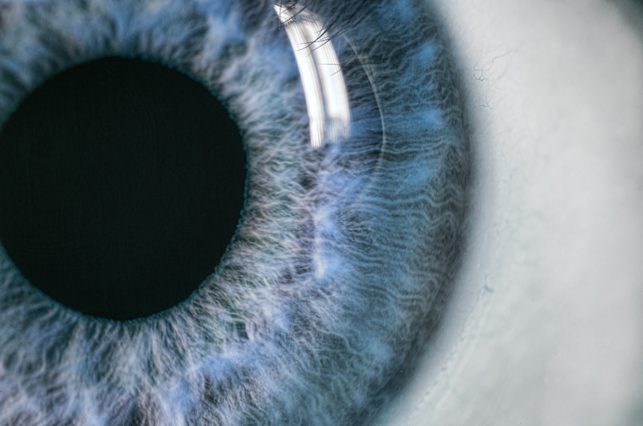
Iris:
The iris is located behind the anterior chamber, which is commonly known as the colored portion of your eye. Inside the iris is the black part in the middle, known as the pupil. The amount of light that gets into your eyes is controlled by the muscles in the iris that constrict and dilate.
Lens:
Right behind your pupil is where the lens is located. The lens changes shape to help your eyes adjust and focus.
Retina:
This is the light-sensitive tissue that lines the back of your eye. Light that passes through the eye via the cornea is what helps us see detail.
Optic Nerve:
The retina sends light through the optic nerve and further to the brain. It is made up of millions of nerve fibers that transmit impulses to our brain, which are responsible for our sight.
20/20 Vision and Lasik Surgery
Only 35% of adults have perfect vision, which is why so many people have to turn to contacts or glasses to correct their vision. However, Lasik surgery has made major advancements and is an excellent option for those who struggle with their eyesight. One of the main benefits of Lasik is that it can permanently improve your vision and get you back to 20/20. Modern medicine has moved toward laser technologies that are highly effective and very safe. The laser removes a defined amount of tissue from your cornea. The new blade-free approach has the approval of the Food and Drug Administration. And doctors have utilized it for nearly two decades. For Orange County iLasik, look no further!
Lasik in Orange County
Dr. Ghosheh at Advanced Eye Medical is the top eye surgeon and Mission Viejo eye doctor. He has performed Lasik procedures for years and helped numerous patients with their vision problems. Depending on what your needs are, his clinic provides a wide variety of services. Dr. Ghosheh focuses on providing patients with the most cutting-edge eye care. His goal remains to ensure he meets every patient’s unique needs. Dr. Ghosheh is a board-certified ophthalmologist and refractive surgery specialist with experience in laser vision correction, cataract surgery, and corneal transplantation. Contact his office today for an appointment!